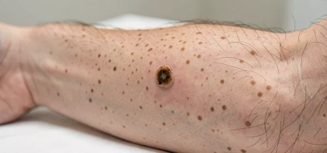
Skin cancers encompass several types of lesions, the appearance of which can vary significantly from one patient to another. Recognizing the visual signs of basal cell carcinoma, squamous cell carcinoma, or melanoma from photos remains a delicate exercise, even for healthcare professionals. The use of images helps familiarize oneself with certain forms, but it never replaces a clinical examination or a biopsy.
Teledermatology and Skin Photography: A Clarifying Framework
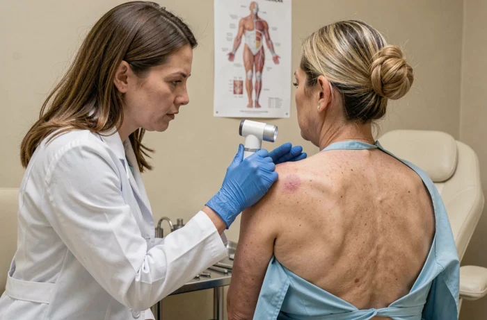
Recent clinical recommendations increasingly emphasize standardized photography for the triage of suspicious lesions. The principle relies on two complementary images: a contextual image showing the affected body area, followed by a close-up of the lesion with a visible scale for measurement. Without these two elements, remote evaluation loses reliability.
This quality requirement sets a clear limit. A photo taken with a smartphone in poor lighting conditions does not provide the same information as a shot taken in a clinic with a dermatoscope. Triage guidelines published in 2026 stress that teledermatology should not replace a physical examination when the level of suspicion is high.
Online resources that compile photos and images of skin cancer help to better understand the diversity of appearances of these lesions. However, the available data do not allow for a conclusion that a self-diagnosis based on image comparison offers sufficient reliability to forgo medical advice.
Visual Signs of Basal Cell Carcinoma and Squamous Cell Carcinoma
Basal cell carcinoma is the most common form of skin cancer. It often manifests as a small translucent or pearly bump, sometimes crossed by visible blood vessels. On certain skin tones, it can take on a pink or brownish hue, complicating visual detection.
This type of carcinoma generally remains localized and does not metastasize. Its growth is slow, but a lesion ignored for months can spread and damage surrounding tissues, including cartilage or bone in advanced cases.
Squamous Cell Carcinoma: A More Aggressive Lesion
Squamous cell carcinoma frequently presents as a crusty wound that does not heal, a firm nodule, or a rough, scaly plaque. It often appears on sun-exposed areas: face, ears, back of the hands, scalp.
Unlike basal cell carcinoma, this cancer can invade lymph nodes and produce metastases. A persistent wound for several weeks in an exposed area warrants an immediate consultation.
- Pearly or translucent bump with visible vessels: consider basal cell carcinoma
- Crusty wound that does not heal after three to four weeks: suspect squamous cell carcinoma
- Red, scaly, rough plaque on an exposed area: consult a dermatologist
- Firm, rapidly growing, sometimes painful nodule: do not wait before seeking medical advice
Melanoma and Suspicious Mole: The ABCDE Rule and Its Limits
The ABCDE rule remains the most widely disseminated guideline for evaluating a mole. Asymmetry, Irregular Borders, Non-homogeneous Color, Diameter greater than six millimeters, Evolution over time. This mnemonic framework is simple, but it does not cover all situations.
Some melanomas develop without presenting all of these criteria. Nodular melanomas, for example, can be symmetrical and uniformly colored while being aggressive. The change in appearance of a lesion is the most reliable warning sign, regardless of its initial appearance.
The Ugly Duckling Sign
This concept usefully complements the ABCDE rule. It is based on comparative observation: among all the moles of a person, the one that does not resemble any other deserves special attention. A mole that clearly stands out from its neighbors should be examined by a specialist.
Field reports vary on the sensitivity of this sign according to phototypes. On darker skin, melanomas more often develop in areas less exposed to the sun (palms, soles of the feet, under the nails), making visual comparison with other moles less relevant.

Monthly Skin Self-Examination: Conditions and Protocol
Updated recommendations from the American Cancer Society in August 2026 detail a protocol for monthly self-examination. The examination should take place in a well-lit area, preferably in natural light, with a full-length mirror and a handheld mirror to inspect hard-to-reach areas.
Assistance from a loved one is recommended to examine the scalp and back. These areas represent common blind spots during self-examination. Reviewing each part of the body systematically (face, neck, torso, arms, legs, feet, spaces between toes) increases the chances of spotting an abnormal lesion.
- Photograph existing lesions to track their evolution from month to month
- Do not let taking photos delay a consultation if a lesion appears suspicious
- Report any new spots, or any changes in size, shape, or color to a doctor
Self-examination does not replace professional screening. For individuals at high risk (personal or family history of skin cancer, numerous atypical moles, light phototype), regular dermatological follow-up remains the standard. Detecting melanoma at an early stage significantly improves prognosis, justifying the combination of rigorous personal monitoring and appropriate medical follow-up.